Forward self-dimer
Risk: Low
- Longest run
- 4 bp
- 3′ run
- 1 bp
- GC pairs
- 2
- Approx. ΔG
- -7.0
Check a forward and reverse primer pair for self-dimers, hetero-dimers, 3′ complementarity, GC content, and Tm balance. Use Basic mode for a fast screen. Use Advanced mode when primer concentration, salt, or qPCR sensitivity changes the risk.
Paste primers in 5′ to 3′ orientation. The calculator scans anti-parallel complementarity and flags runs that can produce primer-dimer bands or qPCR melt peaks.
Basic mode screens a primer pair fast. Advanced mode adds concentration, salt, alignment, and assay warnings.
Paste the forward primer in 5′ to 3′ orientation. Non-DNA characters are ignored.
Length
20 nt
GC%
45.0%
Tm
44.6°C
Forward primer bases
Paste the reverse primer as ordered from the vendor, also written 5′ to 3′.
Length
20 nt
GC%
50.0%
Tm
46.7°C
Reverse primer bases
Live primer-dimer result
Worst signal: Reverse self-dimer. Longest complementary run is 4 bp, with 4 bp at a 3′ end.
The strongest warning comes from 3′ complementarity. Polymerase can extend this kind of primer-dimer.
Risk: Low
Risk: High
Risk: Moderate
The lower line shows the reverse-complement orientation used for primer pairing.
Query   5′           GCTTACAGGATCGTCAACGA 3′
                     | |  | |
Partner 3′ TCGTTGACGATCCTGTAAGC           5′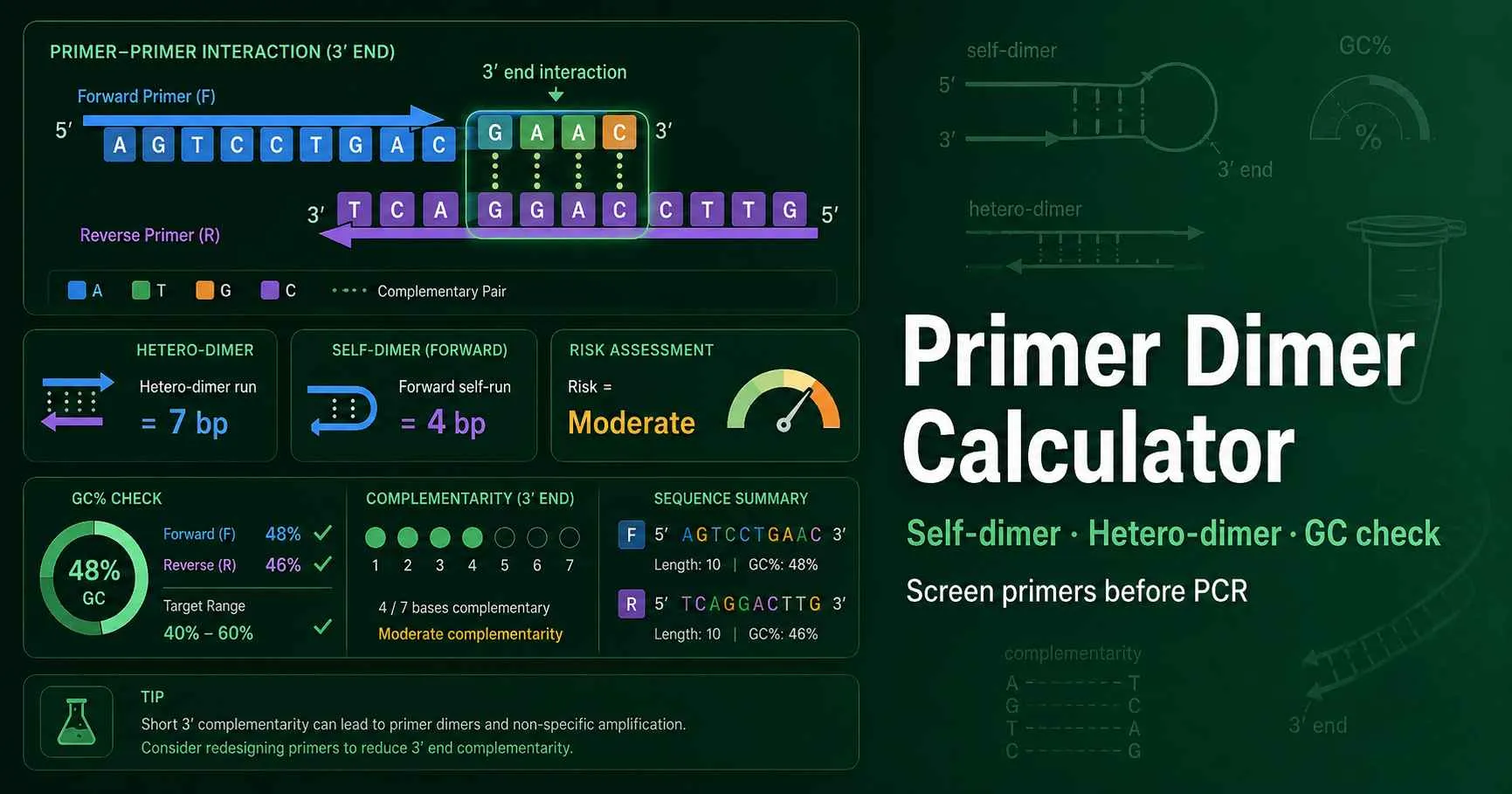
A primer dimer forms when PCR primers anneal to each other instead of the target DNA. The risky version occurs at the 3′ end, because DNA polymerase can extend that paired end. A primer pair can look perfect by length and GC content, yet still fail because the two 3′ tails bind each other.
This calculator tests three events: forward self-dimer, reverse self-dimer, and forward-reverse hetero-dimer. It also reports GC%, approximate Tm, 3′ clamp behavior, and the best alignment that explains the warning. IDT’s oligo-analysis workflow also separates hairpin, self-dimer, and hetero-dimer checks, which matches the same design logic used here. Review IDT OligoAnalyzer features.
Use the result as a screening step before ordering primers or setting up qPCR. A low-risk score does not guarantee perfect amplification. A high-risk score tells you to redesign at least one primer before you spend reagents.
Enter the forward primer in 5′ to 3′ orientation. The tool removes spaces and ignores non-DNA characters.
Enter the reverse primer as ordered from the oligo vendor. Do not reverse-complement it before pasting.
Check the longest complementary run and the 3′ complementary run for each primer and primer pair.
Add primer concentration, salt, and amplicon length to judge qPCR or PCR risk more carefully.
Screens one oligo for self-dimer risk and gives length, GC%, and Tm.
Checks the second primer exactly as ordered, written 5′ to 3′.
Shows whether the forward and reverse primers can anneal to each other.
Highlights complementarity that DNA polymerase can extend.
Adds salt, concentration, amplicon length, and alignment display.
The primer pair shows no strong 3′ complementarity. Routine PCR should still include a negative control.
A run of 3–4 bases or a stable internal match may cause faint primer-dimer bands.
A 3′ complementary run can create extendable primer-dimer products. Redesign one primer.
The National Institute of Justice primer-design guidance warns against complementarity between primers, especially at the 3′ end, because it can create amplified primer-dimers and primer-oligomers. Read the primer-complementarity guidance.
A qPCR assay uses a 96 bp amplicon, a 23 nt forward primer, and a 22 nt reverse primer. The calculator reports 48–52% GC, a Tm difference below 2°C, and no 3′ run longer than 2 bp. That pair deserves a low-risk label.
You would still run a no-template control. If the melt curve shows one target peak and no early NTC amplification, the primer design likely supports reliable relative expression analysis.
A second primer pair shows a 4 bp complementary run at the reverse primer 3′ end. The run contains two G·C pairs, so the small duplex can hold long enough for extension. The calculator labels the pair moderate or high risk, depending on the full alignment.
Redesign the reverse primer first. Shift it three to five bases when the target region allows it. Then check the new pair with the DNA melting temperature calculator before setting annealing temperature.
Start with target specificity, then check primer physics. Choose primers that match the target locus, avoid long homopolymer runs, keep GC content near the usual 40–60% range, and keep Tm values close. After that, run dimer and hairpin screens.
A primer pair that passes this page should still fit the full PCR setup. Use the PCR master mix calculator when you need final primer concentration and reaction-volume planning. Use the hairpin calculator when one primer contains internal complementarity that may form a stem-loop.
Check intramolecular stem-loop structures in the same oligo before ordering primers.
Estimate primer Tm from sequence and reaction conditions before choosing an annealing temperature.
Plan reaction volumes after you choose primer concentrations and final PCR setup.
A primer dimer calculator checks whether one primer can bind to itself or to the other primer in a PCR pair. The most important warning comes from complementarity at the 3′ end. DNA polymerase can extend a paired 3′ end, so a short 3′ dimer can create amplified primer-dimer products. This calculator reports self-dimer risk, hetero-dimer risk, longest complementary run, 3′ run length, GC content, and approximate Tm balance.
Four or more complementary bases at a primer 3′ end deserve attention. Three bases can still matter when the run contains several G·C pairs or when primer concentration is high. Long internal runs also matter because they can stabilize primer pairing before the 3′ end aligns. Treat the result as a design screen, then confirm with a no-template control and melt-curve data for qPCR.
DNA polymerase extends from the free 3′ hydroxyl group. If two primers bind at their 3′ ends, polymerase can copy one primer using the other primer as a short template. That reaction creates a primer-dimer amplicon even when the target DNA is absent. Internal complementarity can still reduce efficiency, but it usually causes less trouble than an extendable 3′ dimer.
Yes. SYBR Green and other intercalating dyes detect any double-stranded DNA, including primer-dimer products. A primer dimer can lower Ct values, distort standard curves, and create an extra melt-curve peak. Probe-based qPCR can tolerate some non-specific products better, but primer-dimer formation still consumes primers and polymerase. Always inspect no-template controls when the calculator reports moderate or high risk.
Many PCR primers work best around 40–60% GC content. A primer below that range can anneal weakly, while a very GC-rich primer can form stable secondary structures. The 3′ end often benefits from one or two G or C bases, but too much 3′ GC complementarity between primers can trigger dimers. This calculator checks GC percent and 3′ clamp behavior together.
No. This calculator gives a fast educational screen for self-dimer, hetero-dimer, and 3′ complementarity. Full thermodynamic tools use nearest-neighbor parameters, oligo concentration, salt, magnesium, and modified bases. Use this page to catch obvious problems before you order primers. Use a dedicated oligo-analysis program for final ordering decisions when the assay costs time or money.
Move one primer a few bases upstream or downstream first. That small shift often breaks a 3′ complementary run without changing the amplicon too much. You can also reduce excessive 3′ GC pairing, adjust primer length, or pick a nearby target region. For qPCR, keep the amplicon short and test no-template controls before trusting expression data.